EE/CSE 577 Final Project
Assigned: Nov 1, 2017
Proposal due: Nov 6, 2017
Status reports due: Nov 27, 2017
Posters to Shu to print: Dec 9, 2017 11:59pm in Drop Box
Final poster presentation: Dec 11, 2017
Final reports due: Dec 13, 2017 NOON in Drop Box
Synopsis
For the final project, we anticipate that people
will work in teams of two or three.
There are two options for this project, which will take roughly five weeks:
- Implement a state-of-the-art research paper from a recent computer
vision/ medical imaging conference or journal (MICCAI, EMBC, SPIE, AAPM et
al.).
- Complete a short research project (more fun!).
You can devise your own project from scratch, or use one of the ideas
suggested below. In either case, the purpose is to get a feel for doing research in
medical image analysis.
All projects must work on the medical imaging data introduced in the class.Guidelines
Option 1
Start by searching through recent medical imaging conference proceedings or
journal articles, and choosing a paper that interests you. You should select a
paper that is appropriate for a four-week project. I.e., it should be more
involved than one of the class assignments. Our expectation is that you will
implement the method yourself rather than using any code that the authors
make available .
Option 2
For this option we'd like you to do a research project with some novelty,
i.e., something that no one has published before. Naturally we're not expecting
PhD-level research in this amount of time, but since two or three of you will be
working together you should be able to come up with exciting results :)
You can choose to work on the medical imaging datasets we provided (see
Project ideas for details) or find a dataset for your own project.
Here are some
challenges for medical image analysis.
Following, are some examples of what we have in mind.
- An interesting extension of prior work. In most cases, we'd
recommend implementing the prior method yourself, rather than downloading
implementations available online, as this gives you a better understanding
of how the method works (and you can avoid mucking around with some one
else's code). But this is not a hard and fast rule -- if the extension is
very significant you may use available code.
- A new application of prior work. Apply a known technique to a new
application domain, and evaluate its performance.
- Develop a new solution (hopefully better!) to an existing
problem. If you chose this option, you have to figure out the solution by
the time you submit the proposal, to convince us that your method will work.
- Pose a new technical problem and solve it. Identify a new problem
for which no known solution exists, devise a solution, and implement/test
it. If you chose this option, you have to figure out both the problem and
solution by the time you submit the proposal.
How ambitious/difficult should your project be? Each team member should count
on committing substantially more effort than on the previous class assigments.
Requirements
Proposal
Each team will turn in a one-page proposal describing their project. It
should specify:
- Your team members
- Project goals. Be specific. Describe the input and output.
- Milestones you anticipate reaching along the way.
- Dataset that you want to use.
Each team must submit a proposal, even if you choose one of the research
ideas described below.
Status reports
Each team will post a status report in the middle summarizing progress to date.
Final poster presentation
We will have a poster session a
Dec 11, 2:30 to 4:30 in the EE Atrium, where each group will present their project to the class. Details will be announced closer to the time of the
poster session.
Final Write-up
Turn in a writeup paper (by noon, Dec 13) describing your
problem and approach. It should include the following:
- Title and team members
- Short intro
- Related work, with references to papers, web pages
- Technical description including algorithm
- Experimental results
- Discussion of results, strengths/weaknesses, what worked, what didn't.
- Future work and what you would do if you had more time
Turn in format
All the writeups should be submitted through Catalyst dropbox and in the NIPS format.
Project Ideas for Option 2
Here is an overview of
all challenges that have been organized within the area of medical image
analysis. There are tons of medical image datasets for you to download. Try to
find an interesting one, and start a research project with it.We also provide several medical imaging datasets and possible project ideas.
Human subject training
HSD is required to access the datasets. Please email Shu
liangshu@cs.washington.edu with
the certificate if you want to use the data. Feel free to use the dataset and choose variations of the ideas or to devise
your own research problems that are not on this list. You can either leverage
machine learning or not, depending on your skill set and target. We're happy to
meet with you to discuss any of these (or other) project ideas in more detail.

1. 3dMD human head dataset. The database includes meshes of 1204 distinct Caucasian individuals, ages
3-40 obtained by a 3dMD digital stereophotogrammetry system. The database does
not include texture or color information. Each mesh includes 15K-20K vertices.
Subjects all face forward, have a neutral expression, and wear caps to remove
hair occlusions. Meshes are cleaned by trained personnel. You can
perform gender or age classification.

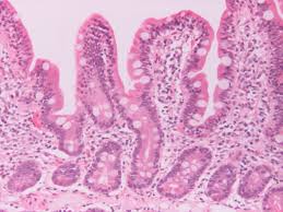
2. Cancer biopsy dataset. The task is to classify a given region of interest (ROI) from a whole slide biopsy to one of the four diagnostic categories: benign, atypia, DCIS and invasive. There are 428 ROIs marked and diagnosed by expert pathologists. ROIs have different sizes and shapes but each has only one diagnostic label. You can use different approaches to overcome size differences: sliding windows, resizing etc.

3. 3D Organ Reconstruction. The task is to reconstruct one of
the online datasets such as
LiTS and try
to reconstruct 3D models of organs such as livers.


